Kendrick Labs, founded in 1987, has over 30 years of experience performing western blotting as a service in combination with our 1D and 2D gel electrophoresis services. Our 2D gel electrophoresis utilizes SDS compatible carrier ampholine tube gels (as opposed to SDS incompatible IPG strips) allowing for superior solubility of protein samples. Western blotting conditions are optimized for every client with a standard turnaround time of 1-2 weeks. Common HRP-labeled secondary antibodies are provided at no additional cost. Pricing and package details are available in our catalog.
Contact us by phone (1-800-462-3417) or e-mail (2d@kendricklabs.com) for a free quote.
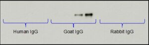
Example Image: Specificity test of our Anti-Goat IgG (H+L)-HRP secondary antibody. No cross-reaction seen with human IgG or rabbit IgG.
Optimized Conditions
With our nearly 40 years of experience, we have optimized protocols and SOPs for gel electrophoresis, transblotting, and western blotting. Antibody specific conditions, such as optimal antibody dilution and blocking reagent, are determined at no additional cost. Records of project details are kept for future work. All client data is strictly confidential.
Detection Capabilities
Kendrick Labs’ experts are experienced at performing chemiluminescent, colorimetric, and fluorescent western blots. Chemiluminescent detection with x-ray film or a CCD camera allows for the highest sensitivity, detecting down to femtogram levels. Fluorescent detection is less sensitive but allows for multiplexing or, the simultaneous detection of multiple protein targets within the same western blot.
Wide Variety of Electrophoresis Options
Kendrick Labs’ specialty is protein gel electrophoresis. We offer a diverse selection of gel electrophoresis options including 1D & 2D gel electrophoresis; reduced, non-reduced, or native gel electrophoresis; and a range of % acrylamide gels to best resolve your protein of interest. Our in-hours gels are poured to two sizes: standard format (13×15 cm) and large format (20×22 cm). We also run a wide variety of commercially available SDS PAGE gels.
Quantitative Western Blotting
Our western blotting services include quantitative western blot analyses. Semi-quantitative western blotting provides a relative comparison between samples on the same western blot. Quantitative western blotting, on the other hand, can determine an absolute amount of a specific protein by loading a standard curve of a purified protein standard.
Reversible Total Protein Stain
Our PVDF membranes are reversibly stained with Coomassie blue prior to western blotting to image the total protein pattern. This image is superimposable with the western blot, allowing for easy matching and excision of proteins of interest from duplicate Coomassie blue-stained gels. The total protein stain also serves to ensure equalized sample loading and transfer efficiency. Other staining options are available upon request.
PTM Blotting Packages
Kendrick Labs offers PhosphoTyrosine, PhosphoSerine/Threonine, and Acetyl-Lysine Western blotting packages at a discounted rate. These packages use optimized in-house antibodies which are included in the price. In a recent pre-print publication Kendrick Labs identified a novel, abundant p-Tyr protein using PhosphoTyrosine Western blotting.
GLP/GMP Capabilities
Our experience with GLP/GMP projects and our robust infrastructure of SOPs set us up to handle your GLP/GMP needs.
Western Blotting Services Overview:
Western blotting, or immunoblotting, is a highly sensitive procedure enabling scientists to use antibodies to detect single proteins or subsets of proteins separated by 1D or 2D gel electrophoresis. For example, antibodies targeting a specific post-translational modification (PTM), such as phosphorylation, can be used to identify proteins with that PTM (phosphotyrosine detection by Western blotting & mass spec citation?). Western blotting can also be useful for monitoring the relative expression of specific proteins, detecting known and unknown proteins in complexes brought down by co-immunoprecipitation, and identifying unknown antigenic targets. After proteins of interest are located by western blotting, corresponding spots can be excised from duplicate Coomassie blue-stained gels and identified by mass spectrometry. In addition, western blotting can also be semi-quantitative and used to determine relative protein expression levels between samples. Special PhosphoTyrosine, PhosphoSerine/Threonine, and Acetyl-Lysine western blot packages using in-house antibodies are available.
Western Blot Methods:
Protein samples are first separated by either 1D or 2D gel electrophoresis, and then proteins are transferred from the acrylamide gel to either nitrocellulose or polyvinylidene difluoride (PVDF) membrane. Membranes are stained with a total protein stain such as Coomassie blue (PVDF) or Ponceau S (nitrocellulose) to image the protein pattern. After staining and imaging, membranes are destained and blocked with a non-reactive protein, such as bovine serum albumin (BSA) or non-fat dry milk (NFDM), to reduce non-specific binding of the antibody. The membranes are then incubated with primary antibody, rinsed to remove excess primary antibody, incubated with a horse radish peroxidase (HRP)-labeled secondary antibody, and rinsed again. Lastly, membranes are incubated in ECL (comprised of luminol and peroxide) where the HRP-labeled secondary catalyzes the reaction between luminol and peroxide producing light. The light emission is detected with x-ray film, and multiple film exposures are provided for each western blot.